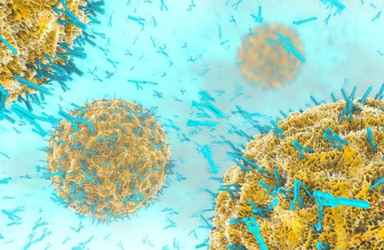
Phage Display vs. Hybridoma: Choosing the Right Method for Monoclonal Antibody Production
Thanks to the advent of methodologies for their identification and development, monoclonal antibodies have revolutionized biomedicine with their applications in disease diagnosis, treatment, and research. Two primary methods dominate monoclonal antibody generation: phage display technology and hybridoma technology.
After you've pinpointed your antigen and are ready to create a new monoclonal antibody for it, familiar questions inevitably come to mind. Should you opt for phage display or stick with the conventional hybridoma method? Many have grappled with the decision, as each technique has its unique strengths and challenges.
This blog dives into the basic concepts of both methods to help you choose the preferred method for your project's needs.
- Understanding Monoclonal Antibodies
- Hybridoma Technology: The Legacy of Milstein and Köhler
- Hybridoma Generation and Selection Process
- Advantages and Strengths of Hybridoma Technology
- Hybridoma Drawbacks and Limitations
- Antibody Phage Display Technology: A Modern Approach
- Antibody Phage Display Library Construction and Screening Processes
- What Are the Advantages of Phage Display Technology?
- What Are the Limitations of Antibody Phage Display Methodology?
- Choosing Between Phage Display vs. Hybridoma for Monoclonal Antibody Production
- Hybridoma vs. Phage Display: The Final Verdict
Understanding Monoclonal Antibodies
Before delving into the methods for their production, it’s crucial to understand what monoclonal antibodies are. Monoclonal antibodies (mAbs) are specialized proteins produced by identical immune cells, all derived from a single parent cell and designed to target specific antigens (foreign substances) in the body. Their high antigen specificity and affinity make them invaluable tools in research, therapeutic, and diagnostic applications.
Each monoclonal antibody consists of two identical heavy chains and two identical light chains, containing constant regions and variable regions. Methods used for monoclonal antibody discovery and production include phage display technology, hybridoma technology, recombinant DNA technology, and single B cell antibody technology.
When deciding between hybridoma development and phage display for monoclonal antibody production, several factors come into play. Let’s explore the key advantages and drawbacks of both techniques to help you determine the best method for your specific needs.
Hybridoma Technology: The Legacy of Milstein and Köhler
Hybridoma technology was pioneered by César Milstein and George Köhler in the 1970s. This traditional method involves fusing immunized animals’ single B cells with myeloma cells (malignant plasma cells) to create hybrid cells capable of producing hybridoma-derived antibodies.
The immortal B cells producing the desired antibody are selected, and monoclonal antibodies with the desired antigen affinity are screened. This intricate cell fusion process, typically triggered by electrical pulses (also known as the B-cell targeting method), PEG, or the pearly chain method, allows these highly efficient mammalian cell lines to be stored long-term for the continuous production of high-quality mAbs.
Hybridoma Generation and Selection Process
- Hybridoma development starts by immunizing animals with the target antigen;
- Single B cells are then isolated from the immunized animal;
- These B cells are fused with myeloma cells to form hybrid cells (hybridomas);
- The hybrid cells are screened and go through a selection process to identify clones producing antibodies with the desired antigen affinity.
Advantages and Strengths of Hybridoma Technology
- Well-suited for assay development due to high affinity and specificity;
- Maintains the natural VH/VL pairing, resulting in increased stability;
- Ability to produce full-length IgG antibodies directly, eliminating the need for additional sequencing, cloning, and transfection into recombinant antibody production systems;
- Large-scale production and high antibody yield;
- Lower cost;
- Mammalian origin minimizes aggregation or recognition failures.
Hybridoma Drawbacks and Limitations
The above-mentioned attributes indicate that hybridomas could be well-adapted for developing therapeutic antibodies. Nonetheless, there are limitations to this method that deserve attention.
- Labor-intensive and time-consuming process (6-8 months on average);
- Use of animals required;
- Murine or animal origin necessitates antibody humanization or antibody chimerization for therapeutic purposes (to avoid immune response), adding extra cost;
- Challenges in humanizing antibodies due to non-human constant regions;
- Genetic instability of cell lines and a risk of cell culture contamination;
- Sometimes difficult and expensive to maintain in culture;
- Gradually being replaced by faster techniques for biotherapeutic development.
Antibody Phage Display Technology: A Modern Approach
Developed by Smith in the 1980s, phage display is a molecular biology technique used to study protein–protein, protein–peptide, and protein–DNA interactions. It leverages bacteriophages (viruses that infect bacteria) to connect proteins with the genetic information that encodes them. Phage genomes are altered, causing the coat proteins of formed virions to bind with other proteins or peptides of interest from any source, presenting them on the surface.
For antibody phage display, instead of peptide antigens, antibodies are displayed on the surface of bacteriophages for the purpose of rapidly identifying antibodies that target a particular antigen. This allows for abundant and rapid generation of antibodies and bypasses the need for animal immunization –it uses phage display libraries to generate antibody fragments with desired properties.
There are four different types of antibody display libraries: immune libraries, naïve libraries, semisynthetic, and synthetic libraries. Libraries can be produced in different formats, namely VHH (from camelid species), Fab (antigen-binding fragment), and scFv (single-chain variable fragment). M13 (E. coli-specific filamentous bacteriophage) and fd filamentous phages are most commonly used, while other phage types, like tailed phages (T4, T7, λ) and icosahedral phages (Qβ and MS2), have also been employed.
Antibody Phage Display Library Construction and Screening Processes
- Antibody genes from individual antibodies are cloned into a phage display vector;
- The vector is used to create a phage display library containing the antibody DNA sequences;
- The library is screened to identify phages displaying antibodies with high antigen affinity.
What Are the Advantages of Phage Display Technology?
Making antibodies by phage display technology has its benefits:
- Large-scale production, high throughput, and rapid screening of diverse clones (with no clone viability issues);
- No need for a host animal and no immunogenicity issues (at least for naïve libraries);
- Quick process (compared to hybridoma); takes a few weeks;
- Possible to directly screen human libraries;
- Suitable for non-immunogenic and toxic antigens;
- Precise control over the selection process;
- Easy to further modify or produce recombinant proteins.
What Are the Limitations of Antibody Phage Display Methodology?
Although an advanced technology, the antibody phage display method has its set of disadvantages, too, like any method:
- Requires careful optimization for specific applications;
- May lack post-translational modifications found in mammalian cells;
- Random pairing of antibody variable regions (VH and VL) and potential loss of natural pairing information;
- Potential for reduced antigen affinity of binders;
- Antibodies might not consistently mimic the in vivo functions or characteristics of naturally existing antibodies;
- Technically challenging, requires expertise in molecular biology and phage handling.
Choosing Between Phage Display vs. Hybridoma for Monoclonal Antibody Production
Each of these two techniques has its strengths and limitations. The answer to the question of which method is best for you lies in your specific goals, needs, and available resources. When deciding between phage display and hybridoma technology for monoclonal antibody production, consider the following factors:
- Project Timeline: If you need antibodies quickly, phage display might be more suitable due to its rapid screening processes. Display libraries are also commercially available.
- Budget: Although less time-consuming, phage display is more expensive than hybridoma.
- Antibody Diversity: For a broader antibody library and antibody diversity, phage display is often preferred since it can screen a larger diversity of antibodies. It’s also better for working with challenging targets.
- Antigen Affinity: If high antigen affinity or high sensitivity is crucial, hybridoma-derived antibodies might be more reliable if you don’t mind the longer generation time.
- Humanization: If you need fully human antibodies, phage display allows easier humanization compared to hybridoma-derived antibodies.
- Expertise: Consider your team’s expertise in molecular biology and cell culture. Each technology requires a specialized set of skills.
Hybridoma vs. Phage Display: The Final Verdict
Both phage display and hybridoma technology have their merits and limitations in monoclonal antibody generation. For projects requiring quick results, broad antibody diversity, and humanized antibodies, phage display often emerges as the preferred method. However, if lower costs or high antigen affinity are your priority, hybridoma technology remains a viable option.
Understanding the basic concepts and strengths of each method will empower you to make an informed decision tailored to your specific needs, whether it’s therapeutic antibodies, research, or diagnostics. Whatever methodology you choose, ProteoGenix offers both phage display and hybridoma development services (and more) that can be tailored and customized to your project’s specific needs.
Feel free to reach out to us if you need more information or expert guidance with your decision. We’re here for you.

