Cart (0 Items)
Your cart is currently empty.
View Products Antibody-drug conjugates
 Thomas Meyer
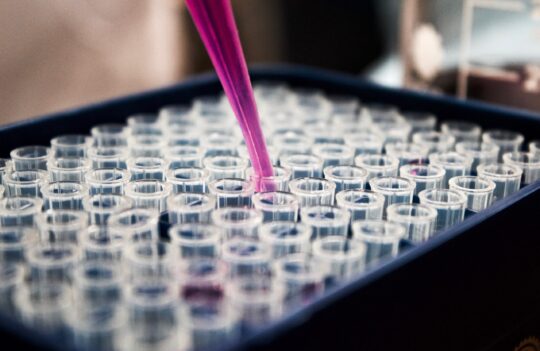
Antibody-drug conjugates (ADCs) represent a powerful class of targeted cancer therapies that combine the specificity of monoclonal antibodies with the potency of cytotoxic drugs.
By selectively delivering highly potent payloads to tumor cells, ADCs aim to maximize antitumor efficacy while minimizing systemic toxicity. Over the past decade, significant technological advances in antibody engineering, linker chemistry, and payload design have accelerated the development of ADCs.
These advances have led to the emergence of multiple generations of antibody drug conjugates, each improving stability, targeting precision, and therapeutic index.
The successful development of ADCs relies on advanced expertise in antibody engineering service, protein production, and conjugation strategies.
An antibody-drug conjugate consists of three key components:
Each component plays a critical role in the overall performance of the ADC.
This modular design has evolved across different generations of antibody drug conjugates, with continuous improvements in each component to enhance efficacy and safety.
The antibody ensures selective targeting of cancer cells, the payload provides cytotoxic activity, and the linker controls drug release within the tumor microenvironment.

Build High-Performance ADCs with Expert Antibody Engineering
Discover our antibody drug conjugate servicesADCs are composed of three main components:
The optimization of these components has been central to the evolution of the different generations of antibody drug conjugates, with each generation improving one or more aspects of structure and function.
The antibody provides targeting specificity, the payload induces cell death, and the linker ensures controlled drug release.
Selecting the right target antigen is essential for ADC efficacy.
Ideal targets:
Advances across the generations of antibody drug conjugates have refined target selection strategies, enabling better tumor specificity and improved clinical outcomes.
The antibody must exhibit:
Later generations of antibody drug conjugates benefit from improved antibody engineering, allowing better control over pharmacokinetics and reduced immunogenicity.
Antibody conjugation methods can be classified as chemical or enzymatic. The chemical conjugation of ADCs depends on the presence and distribution of reactive functional groups on the surface of the antibody. A few examples of the methods that use this type of conjugation include those that leverage lysine (reactive amine groups), cysteine (reaction between cysteine residues and thiol groups inserted in the payload), and tyrosine residues, among others.
In contrast, enzymatic methods rely on the use of enzymes to modify the antibody in a site or sequence-specific manner. The most widely used enzymes in bioconjugation include sortase A (cleaves the threonine-glycine bond and attaches an oligoglycine molecule) and the microbial transglutaminase (modifies conserved glycosylation sites).
One of the most exciting and important trends in antibody design and conjugation is the use of click chemistry for ADC development, also known as bioorthogonal chemistry. These methods rely on very rapid and spontaneous reactions between groups that are not usually found in biomolecules. The term click is used to illustrate the “fusion” between two complementary molecules that can only react with each other. The advantage of this type of conjugation is that is extremely cost-effective, rapid, selective, and it allows better control over the number and distribution of conjugation sites because it does not depend on natural residues present on the antibody carrier.
Want to learn from a real-life project? Discover how we conjugated a client's antibody to be preclinical ready
Read the case reportLinkers can be defined as the interface between biology and chemistry serving as the bridge that links antibodies and drugs in ADCs. Linker chemistry in ADCs dictates the stability, safety, therapeutic potency and window, and efficacy of ADCs. They also determine the mechanism of action of these complex therapeutics, warranting the need for extensive analysis of these drugs before they can be considered ready for clinical studies or trials.
Linker technology can be defined as cleavable or non-cleavable, each catering to different mechanisms of action. Independently of their class, linkers typically consist of two major domains: a payload-binding domain and an antibody-binding domain. Linker optimization has been a key factor in the progression of the different generations of antibody drug conjugates, significantly improving therapeutic index.
Linker chemistry can be divided into cleavable or non-cleavable. However, the vast majority of ADCs in the clinic or in active development can be classified as chemically-labile linkers. These linkers incorporate chemical triggers allowing them to degrade in response to specific signals or conditions. In contrast, non-cleavable linkers are designed to be fully and stably integrated into the payload. In this case, the payload-linker complex is only released upon the degradation of the ADC molecule by lysosomal enzymes.
Generally, cleavable linkers are easier to develop given that they are compatible with a broader range of payloads. Because these linkers are cleaved from the payload, often without leaving residual components, they do not interfere with the drug’s bioactivity. This chemistry enhances the efficiency of drug release by ADCs. Moreover, it is better suited to target solid tumors because it allows the release of payloads in the extracellular space, thus leading to a more efficient uptake by cancer cells that might be devoid of the target antigen (bystander effect). One of the most important drawbacks of these linkers is precisely their sensitivity to chemical stimuli, which might result in the release of payloads off-site and subsequently lead to higher systemic toxicity.
Non-cleavable linkers attempt to overcome these stability issues by ensuring that the drug is only released in the intracellular compartment. This chemistry results in safer and more stable ADCs that can be conjugated to extremely toxic cargoes. However, because these linkers are never cleaved from the payload, they are known to modify its structure, often leading to considerable changes in drug potency. For this reason, payload-drug pairs need to be extensively tested in vitro before developers can estimate their toxicity in vivo. At the moment, very few classes of payloads have been successfully conjugated with non-cleavable linkers.
The vast majority of ADCs in the clinic incorporate peptide linkers such as valine-citrulline (Val-Cit) that are degraded by lysosomal proteases such as cathepsin B. This protease is known to leak to the extracellular compartment in necrotic tissues, resulting in extracellular cleavage of ADCs at tumor sites, especially useful when targeting solid tumors.
Recently, there is a growing interest in the development of alternative linkers such as those containing hydrophilic motifs (e.g., β-glucuronic acid motifs). However, this new generation of linkers has yet to reach its full technological maturity.
Payloads are the bioactive component of ADCs. Most payloads used in ADC generation are too toxic to be administered systemically, for this reason, they can only be used in targeted therapeutic applications. Most cytotoxic agents that have found great clinical success exhibit high activity at the nanomolar or picomolar range. Moreover, these agents must be fairly soluble, non-immunogenic, and possess reactive sites for bioconjugation with a suitable linker.
The majority of payloads used in FDA-approved ADCs can be classified as tubulin inhibitors or DNA-damaging agents. The first class acts on tubulin, leading to the arrest of the growth cycle and subsequent cell death by apoptosis. In contrast, the second class is independent of the growth stage of the cell, alternatively acting as molecular scissors that cleave the DNA leading to cell death.
Due to the progressive increase of the safety of linker chemistry, researchers are actively exploring alternative types of payloads including pyrrolobenzodiazepines (PBD) dimers, which have led to very promising results in short periods. Moreover, constructs that employ the use of payloads such as protein toxins, enzymes, and antibiotics are growing fields of interest with vast potential.
Discover our therapeutic antibody services
| Payload class | Mechanism of action | Examples |
|---|---|---|
| Auristatins | Tubulin polymerase inhibitor | Monomethyl auristatin E (MMAE) and Monomethyl auristatin F (MMAF) |
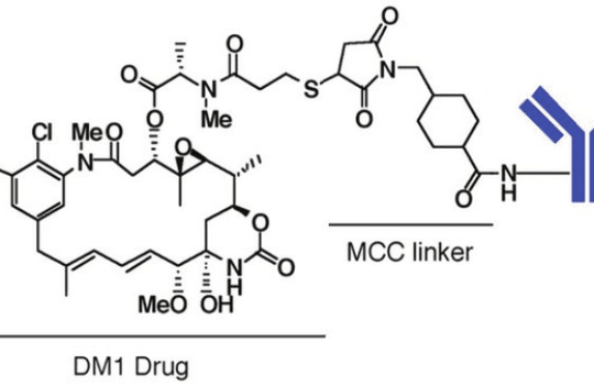
| Maytansinoids | Tubulin depolymerization | Maytansine derivates (DM1 and DM4) |
| Calicheamicins | DNA cleavage | Calicheamicin γ1 |
| Duocarymycins | DNA minor groove alkylating agent | CC-1065 and duocarmycin SA |
| Pyrrolobenzodiazepines (PBD) dimers | DNA minor groove cross-linker | PBD derivates |
| Amatoxins | RNA polymerase II inhibitor | α-Amanitin |
| Protein toxins | Several | Pseudomonas exotoxin (PE) and diphtheria toxin (DT) |
| Antibiotics | Several | 4-dimethylamino piperidino-hydroxybenzoxazino rifamycin (dmDNA31) |
| Enzymes | Several | β-glucuronidase, urease, among others |
You could also be interested in:
Join our email list to receive exclusive content featuring the most interesting industry and research news, biologics development tips pieced together by experts, res, company news, and exclusive limited-offers. Join a community of 80,000 subscribers and save up to 30% on your first order.