

Cart (0 Items)
Your cart is currently empty.
View Products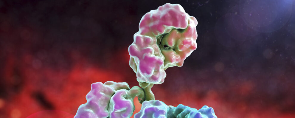 Antibody production
 Thomas Meyer
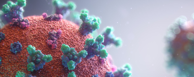
Phage display is a groundbreaking laboratory technique for studying protein interactions with other molecules such as other proteins, peptides, and DNA. The method uses bacteriophages (viruses that infect bacteria) to tether the genetic code with the surface-exposed protein. Here is what that means.
Think of bacteriophages as submicroscopic packages. Inside each package are DNA molecules that encode proteins exposed to the outer surface of the package. Each package, in theory, contains a different DNA sequence that encodes a unique surface-exposed protein. The collection of bacteriophage ‘packages’ are collectively referred to as a phage display library.
Next, the phage-display ‘bait’, is immobilized to capture bacteriophage ‘packages’. The bacteriophages that stick to the bait remain on the surface while other bacteriophages are washed away. The DNA inside the phages is sequenced to reveal the genetic code of the surface-exposed protein that binds the bait. And there you have it. A high-throughput, nonbiased, method for identifying ‘needle in a haystack’-proteins that bind your target of interest.
This technique allows for the screening of large combinatorial libraries, facilitating the identification of peptides, proteins, and antibodies that bind the ‘bait’ with high affinity and specificity. Therefore, phage display allows researchers to link genotype with phenotype, making phage display a versatile tool in molecular biology and biotechnology.
Learn more about ProteoGenix phage display service
Phage display has become a cornerstone in modern biotechnology, thanks to the rapid identification and optimization of high-affinity binders from diverse libraries. Specifically, phage display has accelerated the following biotechnology methods:
This has significant implications for drug discovery, diagnostics, and therapeutic development, where precision and efficiency are paramount. Additionally, phage display technology is instrumental in mapping protein interactions and identifying novel targets for therapeutic intervention.
This blog aims to provide a comprehensive review of phage display technology, elucidating its principles, methodologies, and diverse applications. We will delve into the construction and screening of phage display libraries, explore the different types of libraries, and highlight the latest advancements and trends in the field. By offering detailed insights and practical knowledge, we seek to inform and educate researchers and scientists about the immense potential of phage display technology
Phage display is a molecular technique that involves the expression of peptides, proteins, or antibody fragments on the surface of bacteriophages—viruses that infect bacteria. This method enables high-throughput screening of large combinatorial libraries to identify high-affinity binders. The process begins by inserting a gene encoding a protein or peptide of interest into the phage genome, leading to the display of the encoded protein on the phage’s outer surface. Researchers can then screen these phage-displayed proteins against target molecules to isolate those with the highest affinity and specificity. Phage display technology has revolutionized various fields, including drug discovery, diagnostics, and therapeutic antibody development. By enabling the rapid screening of vast molecular libraries, phage display accelerates the identification and optimization of high-affinity interactions crucial for developing new therapeutics and diagnostic tools.
Phage display technology was first conceptualized and demonstrated by George P. Smith in 1985. Smith’s pioneering work involved the fusion of peptide sequences to the coat protein of filamentous bacteriophage, allowing peptides to be displayed on the surface of the phage particles. This innovation enabled the linkage of the displayed peptide (phenotype) with its encoding genetic material (genotype), facilitating the identification and isolation of peptides with specific binding properties. Smith’s initial experiments used the filamentous phage M13 to display foreign peptides as fusions with the minor coat protein pIII. This groundbreaking approach laid the foundation for subsequent developments in phage display technology, enabling the display of a wide range of peptides and proteins on the surface of bacteriophages.
Phage display technology has continued to evolve, with advancements in library design, selection strategies, and antigen presentation methods. These improvements have enhanced the efficiency and specificity of the technology, enabling the discovery of antibodies and peptides with high affinity and novel functionalities. Today, phage display is a cornerstone of modern biotechnology, widely used in drug discovery, diagnostics, and therapeutic development.
Phage display is a technique where a peptide or protein is genetically fused to a coat protein of a bacteriophage, such that the peptide or protein is displayed on the exterior surface of the phage while the genetic material encoding the protein resides within the phage particle. This allows a direct linkage between the phenotype (displayed protein) and the genotype (encoding DNA). The general workflow of phage display involves the following steps:
Phage display leverages the biology of bacteriophages and their interactions with bacterial hosts. Key scientific principles include:
Phage display libraries are essential tools in biomedical research and drug development, and they can be broadly categorized into peptide libraries and antibody libraries.
The construction of phage display libraries involves several key steps to ensure high diversity and functionality:
Phage display library screening involves several iterative steps to enrich for phages that display peptides or proteins with high affinity for a target molecule:
An immune library is a type of antibody phage display library where the antibody-producing B cells are isolated from an immunized host. Immune libraries therefore contain a heterogenous pool of antibodies that have high affinity toward a particular antigen. The high antibody is a consequence of affinity maturation where B cells or plasma cells have undergone affinity maturation and selection in vivo.
Naive antibody phage display libraries are built from a pool of antibodies that have not been exposed to vaccine antigens and therefore have not undergone any selection or affinity maturation processes. These libraries can be made from humans since naïve libraries do not require host vaccination.
Both naïve and immune antibody phage display libraries can be engineered into different antibody formats by designing PCR primers that bind conserved regions in the desired immunoglobulin heavy or light chains.
Naïve antibody phage display libraries are quicker and cheaper to build compared to immune libraries. Many antibody production companies like ProteoGenix have a diverse collection of pre-built naïve libraries from a range of species and antibody formats.
What immune libraries lack in speed and cost they make up for in precision. Below are the differences between naïve and immune antibody phage display libraries.
| Naive Phage Display Library | Immune Phage Display Library | |
|---|---|---|
| Diversity | High diversity | Limited diversity |
| Affinity Maturation | No | Yes |
| Specifity | +++ | + |
| Library generation time | +++ | ++ |
The numerous applications of phage display technology, in its simplest form, involve identifying proteins that interact with a homogenous immobilized molecule, the bait, with a heterogenous pool of proteins located on the surface of bacteriophages.
Specifically, phage display technology can indiscriminately identify the DNA sequence of proteins, such as small peptide chains or whole antibodies, that bind to any molecule you can dream of with incredible sensitivity. This is a game-changing technology that has revolutionized biomedical science, biotechnology, health care, and the agricultural industry.
This powerful technique has been utilized in various applications, ranging from drug discovery and development to antibody engineering and vaccine design. Let’s explore the wide range of applications where phage display technology.
Phage display technology has revolutionized monoclonal antibody development by allowing the rapid and efficient identification of high-affinity antibodies. This method involves creating vast libraries of antibody fragments, such as single-chain variable fragments (scFvs) or Fab fragments, displayed on the surface of bacteriophages. These libraries are screened against various antigens to isolate antibodies with desired specificities and affinities. Notable monoclonal antibodies discovered using phage display include:
Phage display has also facilitated the engineering of antibodies with enhanced properties such as improved stability, reduced immunogenicity, and enhanced developability, making it a cornerstone in modern therapeutic antibody discovery and development.
Phage display is a critical tool in drug discovery, particularly for identifying peptide and protein ligands that modulate the activity of disease-associated targets. The technology allows for high-throughput screening of phage libraries to identify peptides that bind with high affinity and specificity to target proteins, providing valuable leads for drug development. Key applications in drug discovery include:
The ability to perform iterative rounds of selection and optimization through phage display significantly enhances the efficiency of identifying and refining lead compounds for therapeutic development.
Understanding protein-protein interactions is crucial for elucidating the molecular mechanisms underlying various biological processes and diseases. Phage display technology is widely used to study these interactions by selecting peptides or antibody fragments that bind to specific protein targets. Applications in protein-protein interactions include:
By dissecting these interactions at a molecular level, phage display provides critical information that can be used for drug development and therapeutic intervention.
Phage display has made significant contributions to vaccine development by enabling the identification of immunogenic epitopes. These epitopes can be displayed on the surface of bacteriophages to create phage-based vaccines that elicit strong immune responses. Applications in vaccine development include:
Phage display-derived peptides and proteins are also used as diagnostic tools for detecting immune responses to pathogens, aiding in the development of more effective and safe vaccines.
Phage display allows for the generation of protein variants with altered properties, such as increased stability or binding affinity, which can be useful for developing improved biologics or enzymes. This fascinating phage display application involves making a phage display library with random DNA mutations introduced into the genetic code of a protein of interest. Next, the library would undergo stress such as high temperature, followed by identifying which variants retained the ability to bind its endogenous ligand (the bait).
Phage display can assist in the identification of biomarkers for various diseases, enabling the development of diagnostic tests for early detection and monitoring of conditions. For example, phage libraries displaying human brain proteins have been used to identify patients who present with symptoms of autoimmune encephalitis, a condition where autoantibodies attack the human brain. Here is how it works. The patients who test negative for all brain autoantibodies known to science submit a blood sample to identify and later diagnose, novel autoantibodies causing rare forms of autoimmune encephalitis.
Phage display libraries can be screened against cellular antigens to validate therapeutics. For instance, a newly identified peptide inhibitor can be immobilized and used as bait to identify proteins from cells or tissues that bind to the peptide inhibitor. This approach can also identify undesirable cross-reactive targets of the newly-developed drug.
By displaying random peptide sequences on phages, researchers can screen large libraries for peptides with desired functions, such as receptor ligands or enzyme inhibitors. In this example, the enzyme or receptor is used as bait to identify which peptides the immobilized target interacts with. The DNA encoding the novel peptides can later be used for drug development.
Phage display technology continues to find new applications across various fields of biotechnology and medicine. Some of the emerging applications include:
Phage display technology continues to evolve with advancements in library design, selection strategies, and applications. As the technology matures, it is expected to play an increasingly important role in developing new therapeutics, diagnostics, and research tools.
Library Construction: Phage display technology enables the construction of highly diverse libraries of peptides, proteins, and antibody fragments. This diversity allows for the identification of molecules with high specificity and affinity for a wide range of targets, including proteins, cells, and small molecules.
Customizability: The technology allows for the creation of custom libraries tailored to specific needs, such as human, synthetic, and immune libraries. This flexibility makes it possible to target a variety of antigens and discover unique binding sequences.
Rapid Screening: Phage display accelerates the screening process by allowing high-throughput selection of binding molecules. This is particularly advantageous in drug discovery and therapeutic antibody development, where time is a critical factor.
In Vitro Selection: The entire process can be performed in vitro, eliminating the need for animal models and reducing the time and resources required for discovery and development.
Therapeutic Antibodies: Phage display has been instrumental in developing numerous therapeutic antibodies, such as adalimumab (Humira®) and trastuzumab (Herceptin®), which are used to treat various diseases, including cancer and autoimmune disorders.
Drug Discovery: The technology is widely used to discover peptide and protein ligands that can modulate the activity of therapeutic targets, facilitating the development of novel drugs.
Vaccine Development: Phage display enables the identification of immunogenic epitopes for vaccine development, aiding in the design of effective and safe vaccines.
Affinity Maturation: Phage display allows for the in vitro evolution of molecules through iterative rounds of selection and mutation, leading to the development of high-affinity binders. This process is crucial for optimizing therapeutic antibodies and other biologics.
Functional Studies: The technology is used to study protein-protein interactions, identify functional domains, and understand the molecular basis of binding, providing valuable insights for drug development and therapeutic interventions.
Technical Expertise: Constructing high-quality phage display libraries requires significant technical expertise and resources. The process involves cloning, transformation, and amplification, which can be technically demanding and time-consuming.
Quality Control: Ensuring the diversity and functionality of the library is critical. Poor library quality can lead to reduced success rates in identifying high-affinity binders, necessitating rigorous quality control measures.
Target Presentation: The success of phage display depends on how the target is presented during the panning process. Improper presentation can lead to selection bias, where only certain types of binders are enriched, potentially missing other high-affinity candidates.
Non-Specific Binding: Phage particles can sometimes bind non-specifically to targets or selection surfaces, complicating the identification of true high-affinity binders. This requires careful optimization of selection conditions to minimize non-specific interactions.
Functional Testing: After selecting binders, extensive validation is required to confirm their binding affinity and specificity in functional assays. This step is critical to ensure that the selected molecules are effective in their intended applications.
Downstream Development: Transitioning from phage-displayed binders to fully functional therapeutic molecules involves additional engineering, expression, and purification steps, which can be challenging and resource-intensive.
Patent Landscape: The extensive use of phage display technology in various fields has led to a complex patent landscape. Researchers and companies must navigate intellectual property rights and licensing agreements, which can be restrictive and costly.
Single-chain variable fragments (scFvs) are fusion proteins of the variable heavy (VH) and variable light (VL) chains linked by a short linker peptide and not actually fragments of an antibody. This format retains the antigen-binding specificity of full-length antibodies while being smaller and more stable.
Applications in Research and Therapeutics: scFvs are widely used in research for mapping epitopes and studying protein interactions. In therapeutics, scFvs are utilized in targeted cancer therapies, where their small size allows for better tissue penetration and rapid clearance from the bloodstream, reducing potential side effects.
Go to ProteoGenix scfv discovery service
Fragment antigen binding region (Fab) is the part of the antibody that physically interacts with antigens. They consist of one constant and one variable domain from each of the heavy and light chains of an antibody. They retain full antigen-binding capability but lack the Fc region, making them smaller and less prone to inducing immune responses.
Therapeutic and Diagnostic Applications: Fab fragments are used in therapeutic settings where full-length antibodies are not required, such as in the treatment of macular degeneration (e.g., ranibizumab) or as antivenoms for rattlesnake bites (Digoxin immune Fab and Crofab). These antibody fragments are smaller, easier to penetrate tissues, and are less likely to trigger anaphylaxis because the conserved, non-human, Fc region is removed1. They are also employed in diagnostic assays due to their high specificity and stability.
Go to ProteoGenix Fab phage display screening service
Natural antibodies are bivalent meaning they have two identical antigen-binding sites, while bispecific antibodies are artificial antibodies that can simultaneously bind two different epitopes on the same antigen or from different antigens. These formats are engineered by linking different scFvs or Fabs, often using phage display libraries to select the optimal binders.
Therapeutic Applications: Bivalent and bispecific antibodies are particularly valuable in cancer immunotherapy, where they can bring immune cells into proximity with cancer cells, enhancing the immune response against tumors. They are also used in targeting multiple pathways simultaneously to improve therapeutic outcomes. Currently, three bispecific antibodies are being used in clinical settings. Blinatumomab is utilized to target CD19 and CD3 for the treatment of acute lymphoblastic leukemia (ALL). Emicizumab neutralizes clotting factors IXa and X to treat hemophilia A. Lastly, Amivantamab is a licensed bispecific antibody that is used to treat metastatic non-small cell lung cancer by targeting epidermal growth factor and MET receptors.
Go to ProteoGenix bispecific antibody production service
Nanobodies, or single-domain antibodies (sdAb) an antibody fragments consisting of a single variable domain. With a molecular mass of 12-15 kDa, nanobodies are less than 10% of the mass of human IgG antibodies.
The first single-domain nanobodies were engineered from camelids, referred to as VHH fragments, known for their small, stable, and tissue-penetrating properties compared to other mammalian antibodies. Cartilaginous fish, such as sharks, naturally express exceptionally small antibodies immunoglobulin new age receptor (IgNAR) antibodies. These antibodies can be expressed as even smaller monomeric chains called variable new age receptor (VNAR) fragments for enhanced tissue penetration in vivo.
Applications in Imaging, Research, and Therapeutics: Due to their small size and stability, nanobodies are used in molecular imaging to track disease progression in real time. They are also valuable in research for studying protein interactions and as therapeutic agents for conditions like neurodegenerative diseases and cancer.
Go to ProteoGenix VHH screening service
Antibody-drug conjugates (ADCs) are a class of biological drugs used as cancer immunotherapies. ADCs designed to direct cytotoxic chemical cargo to directly kill tumor cells, unlike chemotherapy which can kill healthy tissues. The antigen-binding region directs the conjugated drug to specific cells, while the cytotoxic agent kills the targeted cells.
Phage display is used to identify high-affinity antibodies specific to tumor-associated antigens, which are then conjugated to cytotoxic drugs to create effective ADCs.
ADCs are currently used in the treatment of various cancers, including breast cancer (e.g., trastuzumab emtansine). Research is ongoing to improve the stability, efficacy, and safety profiles of ADCs.
Go to ProteoGenix antibody drug conjugate services
Role of Phage Display in CAR T cell Development: CAR T cells are engineered T-cells expressing a chimeric antigen receptor that give T cells the new ability to destroy an antigen-specific target cell such as cancer cells. Phage display is used to identify high-affinity antibodies that serve as the binding domain for CAR constructs.
Applications in Cancer Immunotherapy: CAR T cell therapy has shown remarkable success in treating certain types of cancer, such as B cell lymphomas and acute lymphoblastic leukemia (ALL). The ability to target specific cancer cells while sparing normal cells makes this a powerful therapeutic approach.
Phage Display in CAAR Development: CAAR T cells are similar to CAR T cells but instead of being engineered to detect a single antigen expressed on a tumor cell, the T cell expresses a decoy antigen that binds the B cell receptor (BCR), a cell surface-bound immunoglobulin expressed by mature, autoantibody-producing, B-cells. This feature allows T cells the ability to seek and destroy harmful memory B cells, curing the autoimmune condition. Peptide phage display libraries can be used to identify peptide candidates well suited for binding autoreactive BCRs from pathogenic memory B cells.
Multispecific antibodies, also known as bispecific and trispecific antibodies, are engineered to bind multiple epitopes on the same antigen or different target antigens simultaneously, providing broader therapeutic effects. Phage display is a powerful approach to facilitate the identification and optimization of these antibodies to ensure effective binding and functional properties.
Multispecific antibodies are being developed for complex diseases that require targeting multiple pathways, such as cancer and autoimmune diseases. These antibodies offer the potential to enhance therapeutic efficacy and reduce drug resistance.
Use of Smaller Antibody Fragments: Smaller antibody fragments, such as Fabs, scFvs, and nanobodies, are developed for specific therapeutic and diagnostic applications. Their small size enhances solubility while increasing tissue penetration, stability, and ease of production, making them ideal for certain clinical settings.
Development Through Phage Display: Phage display is a key technology for screening and optimizing these fragments, ensuring high specificity and affinity for their targets.
Future Prospects for Therapeutic and Diagnostic Applications: Ongoing research aims to enhance the stability, reduce immunogenicity, and improve the delivery of these antibody fragments to target tissues. Their versatility makes them valuable tools in both therapeutic and diagnostic applications.
Traditional Methods for Creating Antibody Fragments: Non-phage display methods involve cloning the desired section of the monoclonal antibody using polymerase chain reaction (PCR) and inserting the fragment into an expression vector. The expression vector is transfected into cells so it can be converted into antibody fragments that are purified and tested for antigen-binding potential.
This process is time-consuming and tedious process has a major flaw. There is no way of knowing if the fragment will retain the desired antigen affinity, solubility, or stability of the intended application.
The solution is to isolate a series of antibody fragment candidates from an antibody fragment library with high affinity to the target antigen. This increases the odds of identifying at least one high-performing antibody fragment with the desired characteristics.
Summary of Key Points: Phage display is a revolutionary technology that enables the study of protein interactions, the development of monoclonal antibodies, and the discovery of novel therapeutic agents. This technique involves displaying peptides, proteins, or antibody fragments on the surface of bacteriophages, facilitating the high-throughput screening of large combinatorial libraries. Phage display has a wide array of applications, including drug discovery, vaccine development, and the study of protein-protein interactions. The integration of next-generation sequencing and CRISPR-Cas9 technologies has further enhanced the efficiency and precision of phage display, making it an indispensable tool in modern biotechnology.
Final Thoughts on the Importance of Phage Display: Phage display technology has profoundly impacted the fields of molecular biology and biotechnology. Its ability to link phenotype to genotype allows for rapid and efficient identification of high-affinity binders, which is crucial for the development of targeted therapies. Phage display continues to drive innovation in therapeutic antibody development, personalized medicine, and diagnostics. As advancements in this technology continue to emerge, its applications and potential are expanding, promising even greater contributions to science and medicine.
To learn more about how phage display can accelerate your research and development projects, visit our Phage Display Service. Our comprehensive services include antibody library construction, screening, and optimization to meet your specific needs. For more information, feel free to contact us directly. Our team of experts is ready to assist you in harnessing the power of phage display technology to achieve your scientific goals.
Join our email list to receive exclusive content featuring the most interesting industry and research news, biologics development tips pieced together by experts, res, company news, and exclusive limited-offers. Join a community of 80,000 subscribers and save up to 30% on your first order.